The « Quantronic » team from the Condensed State Physics Department published an article in Nature.
The authors :

- Z. WANG
- L. BALEMBOIS
- M. RANČIĆ
- E. BILLAUD
- M. LE DANTEC
- A. FERRIER
- P. GOLDNER
- S. BERTAINA
- T. CHANELIÈRE
- D. ESTEVE
- D. VION
- P. BERTET
- E. FLURIN
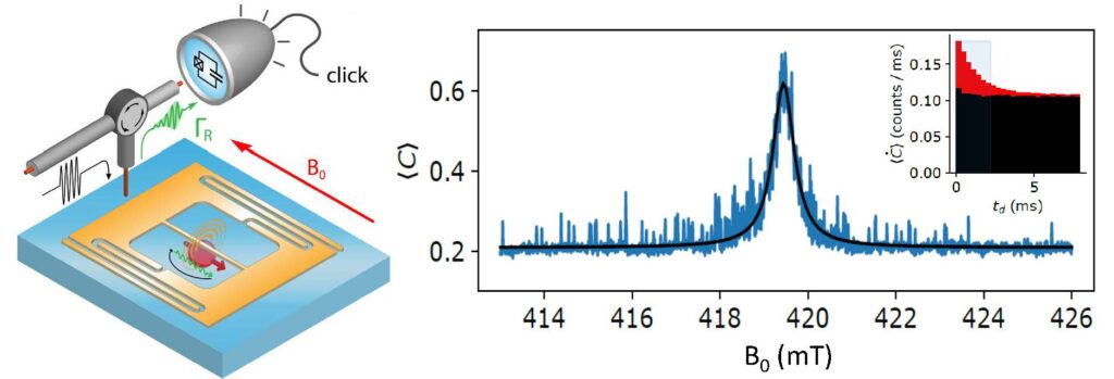
Paramagnetic defects in solids are detected and studied by Electron Paramagnetic Resonance (EPR) spectroscopy, a 80-year old technique. Despite its wide range of applications (chemistry, biology, materials science, …), EPR spectroscopy suffers from a low sensitivity, and can only detect large ensembles of spins, with a spectral resolution limited by inhomogoneous broadening. The Quantronics group at CEA Saclay has recently developed a new method for EPR spectroscopy with single spin sensitivity. The method (described in Fig1 left) consists in coupling magnetically the spin to a planar, micron-scale superconducting microwave resonator, which resonantly enhances its radiative decay rate from the spin excited stae back into its ground state. The spin is excited by a microwave pule, and the microwave photon emitted upon radiative relaxation is detected by a microwave photon counter based on a superconducting qubit, developed in the team. We have demonstrated the method to erbium ions in a scheelite crystal of CaWO4. A typical spin fluorescence curve, showing the count rate as a function of the time after the pulse, is visible in the inset of Fig1 right, showing the spin relaxation in a time ms. Integrating the number of counts for 2ms yields the total number of counts , which is shown as a function of the magnetic field (Figure right). Every time one Erbium ion is resonant with the resonator, a sharp peak is observed. The curve therefore is the first EPR spectrum where the inhomogeneous ensemble line is resolved into its single spin constituents. The method requires low temperatures (10mK), but is applicable to a large class of paramagnetic centers ; it may find applications in EPR spectroscopy as well as quantum computing.

© P. Bertet, Quantronics group, SPEC
Single-spin spectroscopy by microwave photon counting.

